Fájl:Human skeletal muscle tissue 2 - TEM.jpg

Eredeti fájl (2 191 × 1 630 képpont, fájlméret: 1,24 MB, MIME-típus: image/jpeg)
|
Ez a fájl a Wikimedia Commonsból származik. Az alább látható leírás az ottani dokumentációjának másolata. A Commons projekt szabad licencű kép- és multimédiatár. Segíts te is az építésében! |
Összefoglaló
| Leírás |
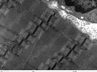
Transmission electron microscope image of a thin longitudinal section cut through an area of human skeletal muscle tissue. Image shows several myofibrils, each with distinct banding pattern of individual sarcomeres. Image of muscle sarcomeres shows distinct banding pattern: the darker bands are called A bands(the A band includes a lighter central zone, called the H band), and the lighter bands are called I bands. Each I band is bisected by a dark transverse line called the Z-line). Paired mitochondria are on either side of the electron opaque Z-line. The Z-Line marks the longitudinal extent of a sarcomere unit. JEOL 100CX TEM |
| Forrás | |
| Szerző | Louisa Howard |
| Engedély (Fájl újrafelhasználása) |
PD |
Licenc
| Louisa Howard, a mű szerzője művét közkinccsé nyilvánította. Ez a világ minden részén érvényes. Egyes országokban ez jogilag nem lehetséges. Ha így van, akkor: Louisa Howard jogot ad bárkinek, hogy bármilyen célból, feltétel nélkül használhassa ezt a fájlt, kivéve a törvény által kötelezően előírt feltételeket.
|
Fájltörténet
Kattints egy időpontra, hogy a fájl akkori állapotát láthasd.
| Dátum/idő | Bélyegkép | Felbontás | Feltöltő | Megjegyzés | |
|---|---|---|---|---|---|
| aktuális | 2006. október 7., 16:58 | | 2 191 × 1 630 (1,24 MB) | Patho | {{Information |Description= Transmission electron microscope image of a thin longitudinal section cut through an area of human skeletal muscle tissue. Image shows several myofibrils, each with distinct banding pattern of individual sarcomeres. Image of |
Fájlhasználat
Az alábbi lap használja ezt a fájlt:
Globális fájlhasználat
A következő wikik használják ezt a fájlt:
- Használata itt: de.wikibooks.org
- Használata itt: fr.wikipedia.org
- Használata itt: ko.wikipedia.org
- Használata itt: lt.wikipedia.org